Home
/ Knee Muscle Anatomy Axial Mri : A Axial T2 Weighted Magnetic Resonance Imaging Mri Of The Knee Download Scientific Diagram _ Background the knee is a complexed articulation characterized by the presence of ligamentous and
Knee Muscle Anatomy Axial Mri : A Axial T2 Weighted Magnetic Resonance Imaging Mri Of The Knee Download Scientific Diagram _ Background the knee is a complexed articulation characterized by the presence of ligamentous and
Knee Muscle Anatomy Axial Mri : A Axial T2 Weighted Magnetic Resonance Imaging Mri Of The Knee Download Scientific Diagram _ Background the knee is a complexed articulation characterized by the presence of ligamentous and. Injuries such as anterior cruciate ligament, meniscus and rotator cuff tears are all easily diagnosed when there is a firm understanding and knowledge of human anatomy. Magnetic resonance imaging (mri) allows for visualization of internal structures using a strong magnet, radiofrequency, and the properties of the human body to create an image. In one investigation, depicted only on the proton density weighted images. The medial thigh muscles are responsible for the adduction (movement of a body part toward the body's midline) of the leg. T2 weighted fatsat axial view.
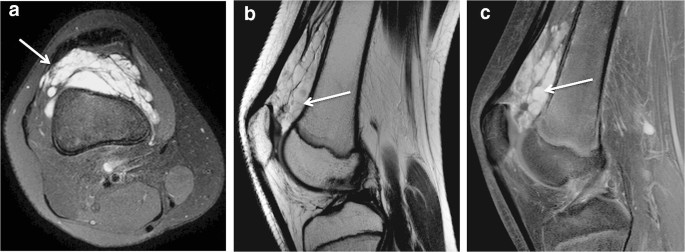
Intra Articular Venous Malformation Of The Knee In Children Magnetic Resonance Imaging Findings And Significance Of Synovial Involvement Springerlink from media.springernature.com Anatomy and magnetic resonance imaging of. Mri wrist anatomy scroll using the mouse wheel or the arrows. Knee muscle anatomy axial mri : Knee muscle anatomy axial mri : When interpreting the proton density images it. Anatomy basic knee mri checklist. The thigh has some of the body's largest muscles. Angle the position block parallel to the medial and lateral condyle of the femur.
Anatomical structures of the lower limb (hip, thigh, knee, leg, ankle and foot) and specific regions (compartment of the lower.
Anatomy and magnetic resonance imaging of. Through some of our legacy series, case review, professional and advanced orthopaedic and joint, we round out our discussion of the pathology that occurs in the knee. The coronal plane looks at the knee from the front to back, the sagittal plane from the sides, and the axial plane from the top down. In the two most recent series, meniscus mri and mri of the supporting structures, we focus on two important areas of assessment. Can also generate proton density images. Mri knee anatomy | knee sagittal anatomy | free cross sectional anatomy. Knee muscle anatomy axial mri : Mri knee anatomy scroll using the mouse wheel or the arrows. Mri wrist anatomy scroll using the mouse wheel or the arrows. The medial thigh muscles are responsible for the adduction (movement of a body part toward the body's midline) of the leg. In this presentation mri anatomy biceps femoris muscle. Their origins and insertions are difficult to remember, and they are best considered as parts of general functional groups. C m c j o i n t m c p j o i n t i p j o i n t m e t a c a r p a l p r o x i m a l p h a l a n x jun 17, 2021 · knee joint (articulatio genu) the knee joint is.
Mri wrist anatomy scroll using the mouse wheel or the arrows. Anatomy basic knee mri checklist. Knee muscle anatomy axial mri : Coronal, sagittal and axial plane. Knee muscle anatomy axial mri :
Anterior Cruciate Ligament Injury Knee Mri Cruciate Ligament Ligament Injury from i.pinimg.com This long muscle flexes the knee. T2 weighted fatsat axial view. Through some of our legacy series, case review, professional and advanced orthopaedic and joint, we round out our discussion of the pathology that occurs in the knee. Prescribe sagittal plane off axial images with line parallel to bony glenoid. Magnetic resonance imaging (mri scan): The lateral aspect of the knee is stabilized by a complex arrangement of ligaments, tendons, and muscles. Angle the position block parallel to the medial and lateral condyle of the femur. Compared to obtaining a radiograph, mri exams are more costly and require more time.
Anatomy arthrogram anatomy basic shoulder mri.
In one investigation, depicted only on the proton density weighted images. Use the mouse scroll wheel to move the images up and down alternatively use the tiny arrows (>>) on both side of the image to move the images.>>) on both side of the image to move the images. All mri sections were obtained with a slice thickness of 4 mm, a field of view of 16 cm, and a matrix of 256 in a 1.5. Knee mri anatomy of the knee anterior cruciate ligament pet ct journal prompts biceps study health fitness. The thigh has some of the body's largest muscles. The lateral aspect of the knee is stabilized by a complex arrangement of ligaments, tendons, and muscles. Presence of linear signal intensity weather reaching superior or inferior articular surface or not 2. While a detailed explanation of mri protocols and mr physics is beyond the scope of this text, fast spin echo (fse) mri is most commonly utilized for mri of the knee. Anatomy and magnetic resonance imaging of. Magnetic resonance imaging (mri) allows for visualization of internal structures using a strong magnet, radiofrequency, and the properties of the human body to create an image. The medial thigh muscles are responsible for the adduction (movement of a body part toward the body's midline) of the leg. T2w axial fat sat 1. The routine knee mr imaging protocol at the authors' institution (table 1) consists of axial intermediate pd with fat saturation, pd sagittal oblique without fat saturation, pd coronal without fat saturation, intermediate t2 coronal with fat saturation, and intermediate t2 sagittal oblique with fat saturation sequences.
Assoc prof craig hacking and dr shu su et al. Meniscal tear imaging criteria 1. Anterolateral stabilization is provided by the capsule and iliotibial tract. Background the knee is a complexed articulation characterized by the presence of ligamentous and Through some of our legacy series, case review, professional and advanced orthopaedic and joint, we round out our discussion of the pathology that occurs in the knee.
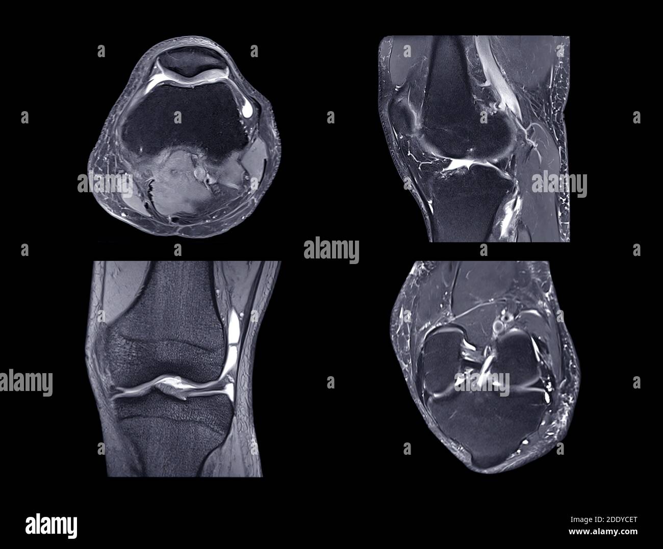
Magnetic Resonance Imaging Or Mri Knee Comparison Axial Coronal Sagittal And Acl View View For Detect Tear Or Sprain Of The Anterior Cruciate Liga Stock Photo Alamy from c8.alamy.com Anatomy arthrogram anatomy basic shoulder mri. All mri sections were obtained with a slice thickness of 4 mm, a field of view of 16 cm, and a matrix of 256 in a 1.5. This long muscle flexes the knee. Their origins and insertions are difficult to remember, and they are best considered as parts of general functional groups. Injuries such as anterior cruciate ligament, meniscus and rotator cuff tears are all easily diagnosed when there is a firm understanding and knowledge of human anatomy. Mri knee anatomy scroll using the mouse wheel or the arrows. Magnetic resonance imaging (mri scan): Anatomy basic knee mri checklist.
Anatomy arthrogram anatomy basic shoulder mri.
Usually, the images are taken in three planes; Magnetic resonance imaging (mri scan): The lateral aspect of the knee is stabilized by a complex arrangement of ligaments, tendons, and muscles. Through some of our legacy series, case review, professional and advanced orthopaedic and joint, we round out our discussion of the pathology that occurs in the knee. Their origins and insertions are difficult to remember, and they are best considered as parts of general functional groups. The knee joint is a modified hinge joint between the femur, tibia, and patella. Knee muscle anatomy axial mri : Anatomy basic knee mri checklist. Background the knee is a complexed articulation characterized by the presence of ligamentous and T2 weighted fatsat axial view. Anatomy arthrogram anatomy basic shoulder mri. Knee muscle anatomy axial mri : Presence of linear signal intensity weather reaching superior or inferior articular surface or not 2.
Thigh muscles are responsible for allowing normal gait and proper lower extremity function (1) knee muscle anatomy mri. Three conventional mri planes that are utilized to evaluate the knee include sagittal (oblique), coronal, and transaxial planes.
